- +971 4 837 83 81
- info@fandascientificme.com
OUR PRODUCTS
FLIMera
SPAD array imaging camera for dynamic FLIM studies at real time video rates
A New Concept in FLIM Imaging
The HORIBA FLIMera camera is a new concept in FLIM technology. It is a wide field imaging camera, rather than a confocal point scanning system, with the intrinsic benefit of being able to study FLIM dynamics at video rates with a simple camera technology.
- TCSPC per pixel architecture offers 24,576 simultaneous decays
- Complex multicomponent lifetimes
- Decay kinetics are acquired directly.
- Sample analysis with up to 5 exponential decays
- EzTime Image simple and efficient software interface
- Perfect for complex and sophisticated molecular analysis like live cell dynamics and detection of biomarkers for disease diagnostics.
Advances in CMOS technology have led to the development of imaging sensors, based on arrays of pixels, with each pixel containing a single-photon avalanche photodiode (SPAD) and its associated timing electronics, based on a time to digital converter (TDC).
This enables rapid (video rate) fluorescence lifetime determination based on the time-correlated single photon counting technique (TCSPC) realized independently in each pixel (Figure 1).
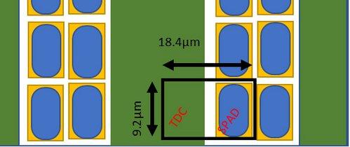
Figure 1: The SPAD and TDC couple
A 192 x 128 pixel image sensor, implemented in 40 nm CMOS technology, is incorporated in a widefield epifluorescence microscope set-up. The sensor has a 13% fill-factor and each 18.4 x 9.2 μm pixel contains a TDC with a resolution <40 ps. This enables up to 24,576 simultaneous fluorescence lifetime measurements each with 4,096 time bins. Through dedicated firmware and the HORIBA EzTime Image software implementation, the fluorescence intensity and the average lifetime as well as the phasor plot generated from the TCSPC imaging measurement, can be simultaneously displayed in real time at video (>30 fps) rates (Figure 2).
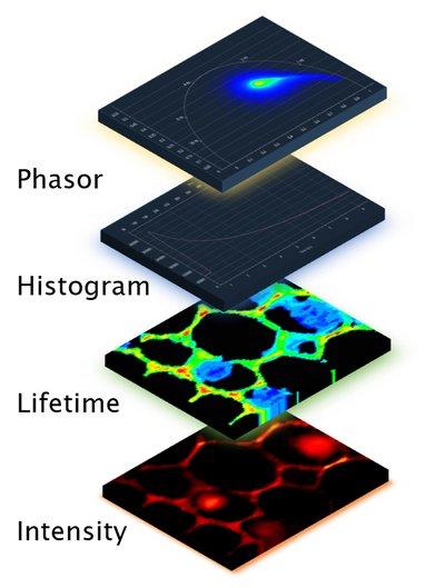
Figure 2: Four simultaneous datasets presented
Modes of operation
The FLIMera is designed for use with HORIBA’s highly intuitive EzTime Image software. This software is used for FLIMera control, data acquisition and analysis. FLIMera provides 4 modes of operation.
- Manual – User start and stop of acquisition, providing TCSPC intensity image and FLIM data for lifetime analysis.
- Timed – User defined run time for acquisition, providing TCSPC intensity image and FLIM data for lifetime analysis.
- Streaming to HDF5 file – TCSPC data can be streamed, for a user-determined time period, to a HDF5 file. This contains full records for each photon in every pixel, so no data is lost, and can be used to reconstruct frames for full decay analysis.
- Phasor Plot – Complementing the histogram representation is the ability to show and select lifetime data in the form of a phasor plot in real time, as seen in Figure 3. The phasor plot requires initial calibration using a lifetime standard.
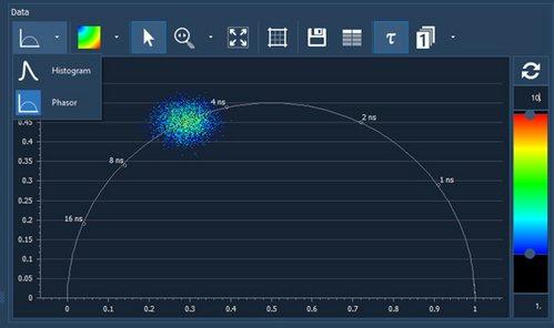
Figure 3: Phasor plot
The video-rate capability is demonstrated using standard samples and FUN-1 labelled yeast cells (Figure 4).
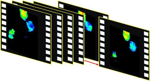
Figure 4: Sequence of images extracted from FUN-1 labeled yeast cell lifetime video
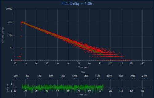
Figure 5 a: Decay from DeltaFLEX cuvette system
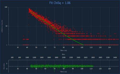
Figure 5 b: Decay from just one of 24,578 SPAD pixels demonstrates the very high resolution and TCSPC fidelity of FLIMera
FLIM and HORIBA

The novelty of the in-pixel detection and timing technology enables a widefield imaging approach, which significantly reduces data acquisition times enabling the study of dynamic events. FLIMera contributes to HORIBA’s long history in the field of fluorescence measurement; recognized by winning the prestigious Institute of Physics Business Innovation Award.
| FLIMera Camera Specifications | |
|---|---|
| Modality | Widefield Time-Domain TCSPC |
| Sensor | SPAD CMOS with In-Pixel TDC |
| Sensor Wavelength Range | 400 to 900 nm |
| Max Total Count Rate | Up to 295 Mcps theoretical |
| Max Pixel Count Rate | 12 kcps |
| Image Array | 192 x 128 pixels |
| Frame Rate (Max.) | 30 fps |
| Fill Factor | 13% |
| Lifetime Range | From 200 ps to 20 ns |
| Lifetime Resolution | Up to 4096 time bins of 50 ps |
| Connection Type | C-Mount |
| Electronics Interface Included | HORIBA FLIMera Base Unit (includes laser synch input TTL) |
| Software Included | HORIBA EzTime Image |
| Required Components | Pulsed laser source |
| Light Source Options | DeltaDiode Lasers |
Similar Products
Related products
-

SA-9600 Series
Learn moreBET Flowing Gas Surface Area Analyzers
-

Aqualog – A-TEEM Industrial QC/QA Analyzer
Learn moreA Simple, Fast, “Column Free” Molecular Fingerprinting Technology
-

Auto SE
Learn moreSpectroscopic Ellipsometer for Simple Thin Film Measurement

